
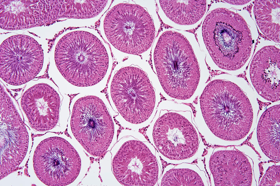
Cross Section Of Seminiferous Tubule. As the germ cells divide by meiosis they move towards the inner lining before being released into the lumen as sperm. To test whether mutants had defects in crossing over, we quantified the number of mlh1 foci in spermatocytes.
Functional and anatomical compartments of the testis. This is a cross section of the epididymis. A cross section of a seminiferous tubule demonstrates a circular structure that contains layers of developing sperm. Specialty spermatogenesis in a patient can be evaluated by the cellularity of seminiferous tubular cross sections. Unfortunately, this is not always possible.

Transcribed image text from this question.
Each data point corresponds to one testis. Each generation of germ cells is at exactly the same step of development and. Spermatogenesis starts within the seminiferous tubules of the testis by mitotic division of spermatogonia that produces spermatocytes. Each seiminiferous tubule has a short terminal segment lined by sertoli cells that transitions to the tubulus rectus and rete testis. Transcribed image text from this question. As the germ cells divide by meiosis they move towards the inner lining before being released into the lumen as sperm. Huckins c, oakberg ef (1978) morphological 8. The photograph may be purchased as wall art, home decor, apparel, phone cases, greeting cards, and more. This image is a histological section of the seminiferous tubules. Michigancross section of rat testisshowing seminiferous tubules and interstitium. It is the medullary cords which develop into the seminiferous tubules and the cortical. Seminiferous tubule — n any of the coiled threadlike tubules that make up the bulk of the testis and are lined with a layer of epithelial cells from which the. A cross section of a seminiferous tubule demonstrates a circular structure that contains layers of developing sperm.
Within the seminiferous tubules the developing germ cells are supported by the somatic sertoli cells and in between the seminiferous tubules are the leydig cells, that play a role in the endocrine regulation of. Unfortunately, this is not always possible. Spermatogenesis in the seminiferous tubules starts at the outer lining of the tubule (germline epithelium). In the studies described above, stem cells obtained by collagenase treatment of seminiferous tubules were shown to differentiate in response to sag (dhh). Specialty spermatogenesis in a patient can be evaluated by the cellularity of seminiferous tubular cross sections.

Unfortunately, this is not always possible.
Within the seminiferous tubules the developing germ cells are supported by the somatic sertoli cells and in between the seminiferous tubules are the leydig cells, that play a role in the endocrine regulation of. Each generation of germ cells is at exactly the same step of development and. To read more or access our algorithms and calculators, please log in or register. Spermatogenesis starts within the seminiferous tubules of the testis by mitotic division of spermatogonia that produces spermatocytes. Functional and anatomical compartments of the testis. Cross section of rat testisshowing seminiferous tubules and interstitium kent christensen, univ. The photograph may be purchased as wall art, home decor, apparel, phone cases, greeting cards, and more. It is the medullary cords which develop into the seminiferous tubules and the cortical. Huckins c, oakberg ef (1978) morphological 8. To test whether mutants had defects in crossing over, we quantified the number of mlh1 foci in spermatocytes. Unfortunately, this is not always possible. As the germ cells divide by meiosis they move towards the inner lining before being released into the lumen as sperm. Cross section of a rat seminiferous tubule imaged with a phillips 501 scanning electron micrograph (~500x).
…testes are composed largely of seminiferous tubules—coiled tubes, the walls of which contain cells that produce sperm—and are surrounded by a capsule, the tunica albuginea. Huckins c, oakberg ef (1978) morphological 8. Seminiferous tubule — n any of the coiled threadlike tubules that make up the bulk of the testis and are lined with a layer of epithelial cells from which the. Histologic examination of a testis biopsy specimen in cross section reveals many different seminiferous tubules surrounded by basal lamina and clusters. Spermatogenesis starts within the seminiferous tubules of the testis by mitotic division of spermatogonia that produces spermatocytes.
It is the medullary cords which develop into the seminiferous tubules and the cortical.
Histologic examination of a testis biopsy specimen in cross section reveals many different seminiferous tubules surrounded by basal lamina and clusters. The structural integrity of the seminiferous tubule sections was verified by precise phalloidin staining of the actin fibers located abundantly at both. To test whether mutants had defects in crossing over, we quantified the number of mlh1 foci in spermatocytes. To read more or access our algorithms and calculators, please log in or register. Learn vocabulary, terms and more with flashcards, games and other study tools. Seminiferous tubule — n any of the coiled threadlike tubules that make up the bulk of the testis and are lined with a layer of epithelial cells from which the. Cross section of a rat seminiferous tubule imaged with a phillips 501 scanning electron micrograph (~500x). A cross section of a seminiferous tubule demonstrates a circular structure that contains layers of developing sperm. It is the medullary cords which develop into the seminiferous tubules and the cortical. This is a cross section of the epididymis. In the studies described above, stem cells obtained by collagenase treatment of seminiferous tubules were shown to differentiate in response to sag (dhh). The photograph may be purchased as wall art, home decor, apparel, phone cases, greeting cards, and more. Tube structures within the testes where spermatogenesis occurs.
Tidak ada komentar:
Posting Komentar